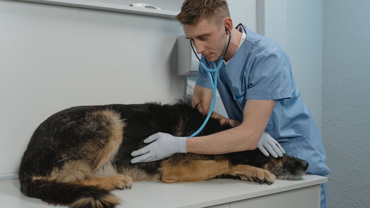
Most tube problems in dogs start after intubation, not during it. A tube that was perfect at induction can slide into a bronchus when the neck flexes, back out when the head turns, or get pinched by a tie pulled too hard. Knowing how to tie in an endotracheal tube in a dog, and checking it afterwards, takes care of most of that.
This guide covers the dog-specific technique for the endotracheal tube (ET tube): pre-measuring, a numbered tie-in sequence, choosing the tie point, breed adjustments and untying for extubation. For principles across species, see our pillar on how to secure an endotracheal tube in dogs and cats.
Quick answer
Pre-measure the tube from the incisors to the thoracic inlet. After intubating and confirming placement, tie a snug clove hitch around the tube just behind the connector, run both ends caudal to the canines, then tie a quick-release bow over the upper jaw, the lower jaw or behind the head. Check the lumen for kinks and keep the pilot line free.
Before you tie: pre-measure the tube length
Lay the chosen tube along the side of the dog's head and neck before induction. The tip should sit at about the thoracic inlet or point of the shoulder, with the connector at the incisor arcade. Mark that point with a pen or tape.
A tube running well past the incisors adds dead space and can slip into a mainstem bronchus. One that is too short can back out of the larynx with little head movement. If you cut a long tube down, push the connector firmly back in.
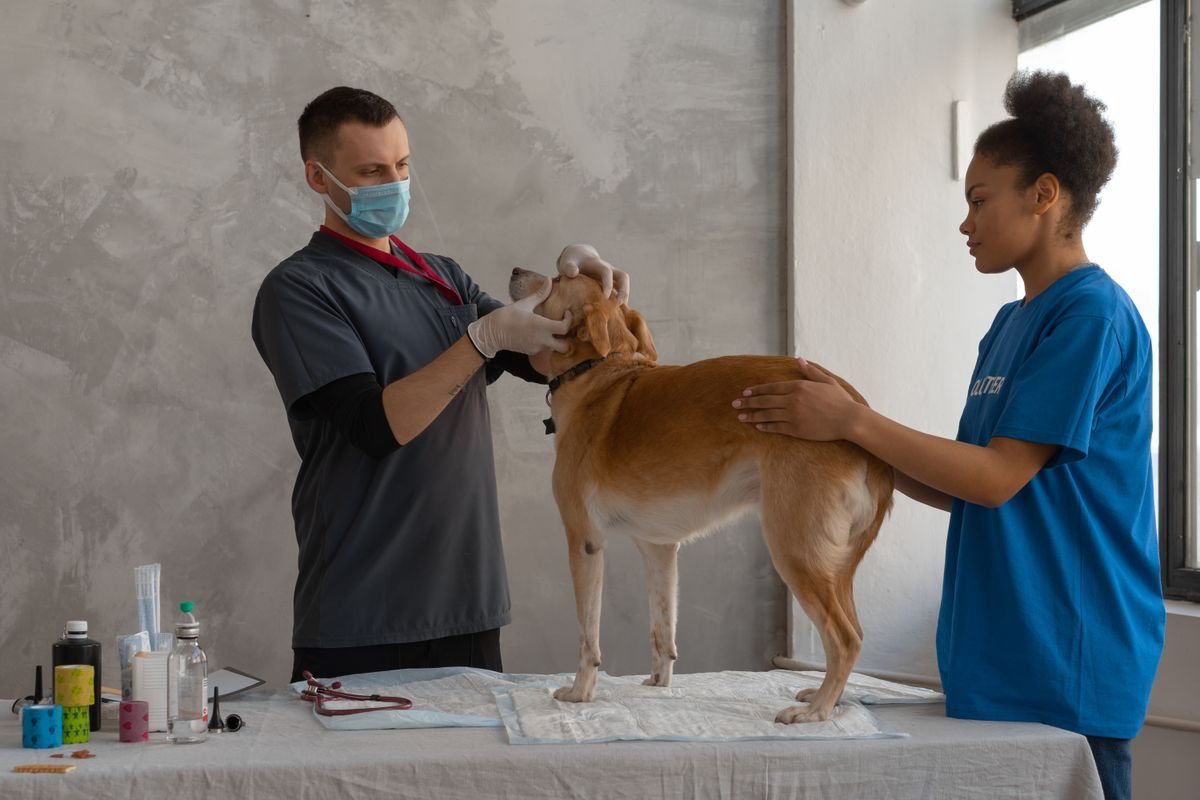
How to tie in an endotracheal tube in a dog, step by step
The usual order is insert, confirm placement, tie in, connect the breathing system, then inflate the cuff, as set out in the University of Bristol canine endotracheal intubation clinical skills guide. Here is the tie-in itself.
- Confirm placement. See the tube between the arytenoids, then look for a capnograph trace and condensation.
- Check your mark. The pre-measured mark should sit at the incisors. Adjust depth now, not after tying.
- Wrap the tube. Form a clove hitch with the middle of the tie just caudal to the connector. Snug it until it stops sliding, without dimpling the tube wall.
- Keep the pilot line out. The pilot line and balloon run outside the wrap, never under it.
- Pass the ends behind the canines. The canine teeth then act as an anchor.
- Choose the tie point. Over the maxilla, under the mandible, or behind the ears (see the table below).
- Finish with a quick-release bow. Never a square knot, so one pull frees the tube in an emergency.
- Connect, inflate and recheck. Attach the circuit, inflate the cuff to just seal, then run the kink check below.
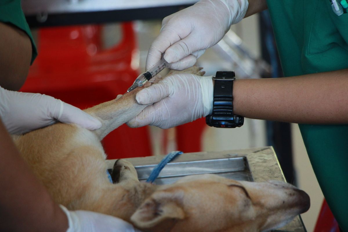
Tie the ET tube to the upper or lower jaw, or behind the head?
Tie away from the surgical field, onto whatever will not move when the head is positioned. Most dogs do well tied over the maxilla, because the upper jaw stays fixed when the mouth opens.
| Tie point | Best for skull type | Best for procedure | Watch for |
|---|---|---|---|
| Upper jaw (maxilla) | Mesocephalic dogs with a defined stop | Most soft tissue, orthopedic and abdominal surgery | Gets in the way of maxillary dental and nasal work |
| Lower jaw (mandible) | Mesocephalic and larger dogs | Maxillary dentals, rhinoscopy, upper oral surgery | Tube moves whenever the mouth opens or closes |
| Behind the head | Brachycephalic and dolichocephalic dogs, toy breeds | Full-mouth oral work, frequent repositioning, CT and MRI | Ear and occipital surgery; excess tension pulls the tube |
For dentals, the tie usually goes to the jaw not being worked on and may need moving partway through. Our guide to securing the ET tube for veterinary dental procedures covers that in detail.
Breed and size differences when securing an ET tube in a dog
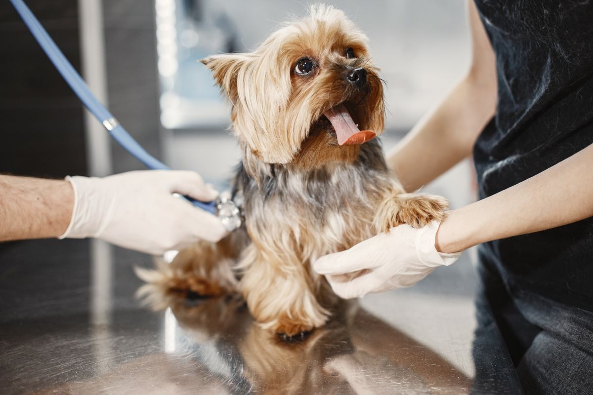
Toy breeds
Small tubes have thin walls and a narrow lumen, so tension that is harmless on a 10 mm tube can narrow a 3 mm tube. Tie just firm enough to stop sliding. Tiny canines give little purchase, so a behind-the-head tie is often easier.
Giant breeds
Heavy heads drag on the tube with every move. Use a tie long enough to wrap generously, finished over the maxilla or behind the head. Recheck depth after moving the dog onto the table.
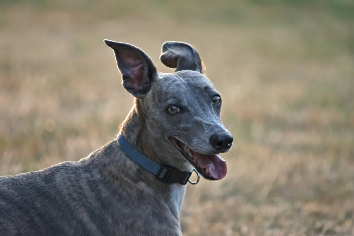
Dolichocephalic dogs
Greyhounds, Borzois and Collies have long, tapered muzzles with no stop, so a tie over the nose tends to slide forward. Bristol's guide mentions taking the ends over the muzzle in long-nosed breeds, but many clinicians find behind the head more stable.
Brachycephalic dogs
There is almost no muzzle to tie to, so tie behind the head. These dogs often need a smaller tube than expected, and the tube usually stays in until the swallow is strong.
Check for kinking or constriction from the tie
Once tied, run a short check before draping.
- Look for dimpling or flattening under the wrap.
- Run a gloved finger along the oral section to feel for kinks.
- Watch the capnograph. A sloping upstroke or rising peak pressure can mean a narrowed lumen.
- Squeeze the reservoir bag. New resistance points to the tie or a kink.
- Confirm the pilot balloon still inflates and deflates freely.
Material matters too. Wet gauze soaks up saliva and can be hard to untie, which we cover in gauze vs purpose-made trach-tube ties. For a broader look, see every ET tube tie option compared.
Repositioning without losing the tube
Before rolling a dog to lateral or dorsal, disconnect the circuit or have someone support it. Move the head and neck as one unit with the body. Afterwards, recheck the depth mark, tie tension and capnograph trace.
If the tie needs moving to the other jaw, keep a hand on the tube at the incisors and re-tie before letting go.
Untying for extubation
Untie once the dog is off gas, so the tube can come out on the first swallow. Keep scissors and a syringe close.
- Pull the bow to release the tie from the jaw or head.
- Deflate the cuff fully and confirm the pilot balloon is flat.
- Slide the tie off the tube, or cut it if it has tightened.
- Extubate smoothly on a swallow.
Trinity disposable ET tube ties were developed by Dr. Billy Davis DVM from more than 23 years in small animal and exotic practice. They are highly visible, do not cling to muzzle hair or soak up saliva, and hold fast to the tube to reduce tube movement.
FAQ
Should I tie the ET tube to the upper or lower jaw in a dog?
The upper jaw suits most procedures because the maxilla stays still when the mouth opens. Use the lower jaw for maxillary dentals and nasal work, and tie behind the head for brachycephalic, dolichocephalic and toy dogs.
Why tie the ET tube behind the canines?
The canines act as a fixed anchor. A tie running caudal to them cannot slide forward off the jaw, so the tube stays at its measured depth.
How tight should the tie be on the tube?
Tight enough that the tube does not slide through the wrap, but not so tight that the wall dimples. Small tubes narrow at much lower tension than large ones.
Why won't the cuff deflate before extubation?
Check whether the tie has trapped the pilot line. Keep the pilot line outside the wrap from the start.
For feline technique, see securing an ET tube in a cat, or browse all our ET tube guides and comparisons. Check our bulk pricing for trach-tube ties, or contact us for phone and fax orders.